Title

Democratizing Discovery: Seeing Diseases Through PRISMS

Subhead
The Coskun Lab makes mapping cells cheaper, customizable, and easier to access.
ID
Dec 02, 2025 | By Leeanna Allen
News Image
A person wearing a white lab coat and blue gloves is working at a laboratory station equipped with advanced imaging equipment. The setup includes multiple tubes, wires, and containers connected to a black optical device. A large monitor on the right displays a vivid, high-resolution microscopic image in shades of purple and pink, showing tissue structures.
Image Caption
Ph.D. student Nicholas Zhang uses PRISMS to generate a high-resolution multiplex image of stem cells.
Widgets

Understanding diseases means knowing not only how cells and molecules function in the body, but where they are located. Spatial omics combines imaging with biological data to map these positions so that researchers can learn how diseases and treatments affect cells within their environment.

Creating these detailed maps is not easy. Instrument and operating costs average $500,000 but can top $1 million for the complex imaging and analysis. Systems rarely work together, and sample preparation takes time, space, and specialized expertise.

Those requirements put the technology out of reach for many labs. 

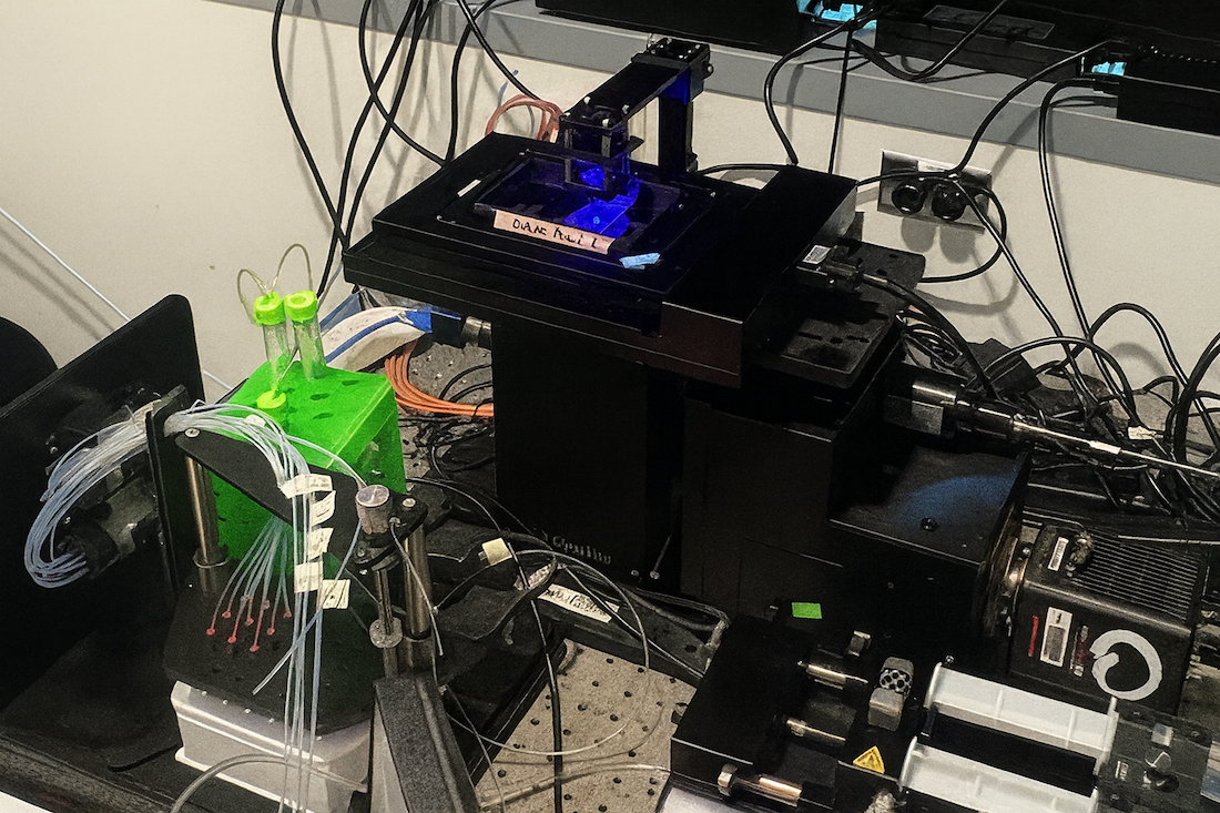
Biomedical engineers at Georgia Tech and Emory University set out to remove these roadblocks to make the technology cheaper, customizable, and easier to access, while still producing high-quality datasets. They created Python-based robotic imaging and staining for modular spatial omics (PRISMS), an open-source, automated process, and recently published in the journal Lab on a Chip. 

With PRISMS, labs can map large areas of tissue as well as zoom in on proteins and biomarkers at 10%-20% of the usual cost. The modular nature means researchers can mix and match components and capabilities according to their needs and even add in their own algorithms.

Ahmet Coskun and his team developed PRISMS using a flexible, modular approach, combining off-the-shelf lenses, lasers, and reagents. Ph.D. student Nicholas Zhang created open-source algorithms and training videos to help users navigate the platform. Automated sample preparation workflows cut costs even more. These cost-savings can allow labs to run more samples, analyze larger datasets, and better collaborate with other labs.

"People can bring their questions, samples, and assays and easily adapt them to PRISMS," Coskun, Associate Professor of Biomedical Engineering at the Wallace H. Coulter Department of Biomedical Engineering, said. “It’s a service to the community for us to help bring spatial omics to more people. That’s why we’re passionate about sharing this with other researchers." 

Close-up of a laboratory imaging setup with a black optical device holding a sample under blue light. Several transparent tubes connect to a green holder with capped vials, alongside pumps and wiring on a perforated metal surface.
PRISMS is compatible with a variety of formats, samples, and processes. This photo shows an example of an imaging setup.

Like any open-source system, there are lots of moving parts and finding the right sequence of steps was a challenge, Coskun said. The team tested different sequences to optimize workflows and the programming language for different tasks. These iterations not only served to improve PRISMS, but also sparked ideas for new modules, like options for temperature control. 

Now, the team is applying spatial omics to look at how diseases from cancer to cystic fibrosis develop in the body and find new treatment targets. Future activities include using PRISMS to increase the capacity of his lab to collaborate with other researchers. 

Coskun notes that Coulter BME is the ideal place to develop technology like PRISMS, “Our students have the engineering expertise to put together these gadgets and do-it-yourself modular systems.”


Citation

Zhang N, Fang Z, Kadakia P, Guo J, Vijay D, Thapa M, Dembowitz S, Grakoui A, Coskun AF. Modular, open-sourced multiplexing for democratizing spatial omics. Lab Chip. 2025 Sep 8;25(25):5379-5392. doi:10.1039/D5LC00286A.

About the Research

This research was supported by the National Science Foundation, grant No. 2338935 and the National Institutes of Health, grant Nos. R35GM151028, T32GM142616, and 1R21AI173900. Any opinions, findings, and conclusions or recommendations expressed in this material are those of the authors and do not necessarily reflect the views of any funding agency. 

Media Contact

Contact the BME Communications team to connect with a faculty member or student about academics or research happening in the Wallace H. Coulter Department of Biomedical Engineering.