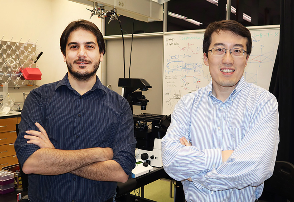
A team of Georgia Tech and Emory researchers has introduced a revolutionary system to enhance the clarity and quality of fluorescence microscopy images. Their innovation, Multiscale Image Restoration through Optimally-Sparse Representation, or MIRO, could transform the way scientists view and interpret microscopic images.
“This has the potential to expand the horizons of biological knowledge,” said Shu Jia, professor in the Wallace H. Coulter Department of Biomedical Engineering at Georgia Tech and Emory, who led the research team. Their groundbreaking study is published this month as the cover story in the journal Science Advances.
Fluorescence microscopy stands as an unrivaled tool in biological research, illuminating the depths and functions of cellular structures. But Jia stressed the need for improved image clarity.
“The inherent limitations of current imaging detectors have posed significant challenges, with various noise sources clouding the resultant image,” he said.
Such noise impedes the precise observation of fluorescent signals, thereby restricting the full potential of fluorescence microscopy.
“The sensitivity discrepancies of current sensors coupled with pixel-to-pixel variations result in uncertain photon detection,” noted the study’s lead author, Biagio Mandracchia, a former postdoc in the Jia lab and currently an independent researcher at the University de Valladolid in Spain. “This has led to the production of images marred by noise, leaving researchers with suboptimal visual data for analysis."
A Clearer Future
The research team believes MIRO represents the future of image restoration.
“MIRO offers a comprehensive algorithm to conquer all noise sources pertinent to microscopy,” said Jia, whose collaborators include Shuichi Takayama, professor in the Coulter Department, as well as Shuyi Nie from Georgia Tech’s School of Biological Sciences, and Chunhui Xu, professor in the Department of Pediatrics at Emory’s School of Medicine.