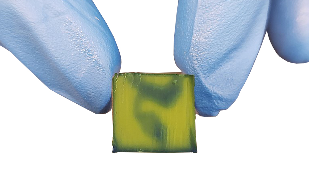
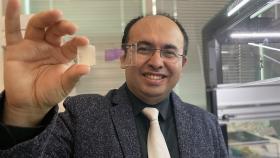
A bioprinted embryonic human heart model at 84 times the actual size. This is one of two different kinds of models Vahid Serpooshan is developing that are 3D printed using soft, flexible hydrogel materials infused with cells from specific patients. Funded by a National Science Foundation Early Career Development award, the models will mimic the exact structure of the heart at two stages: the embryonic heart tube present at roughly 20 days after conception and a more fully developed fetal heart at 30-34 weeks. (Photo Courtesy: Vahid Serpooshan)
When babies are born with severe heart defects like pulmonary artery atresia or hypoplastic left heart syndrome, the prognosis is difficult. There is no cure, no reliable therapy for many of these defects. Just uncertainty. And drastic efforts to fix the parts of the heart that didn’t develop properly.
Ultimately, these tiny babies may face multiple significant surgeries in their early weeks of life.
That’s what Vahid Serpooshan thinks about when he’s in his lab using a sophisticated 3D bioprinter to create models of the earliest stages of heart development: the babies and their families and how his team can help by unravelling some of the mysteries of the developing human heart.
“These are babies who are a few days old and who are suffering from very severe, acute heart disease and heart defects. And many of them do not survive — even after multiple surgeries,” Serpooshan said. “Being able to simulate such severe situations in bioprinted and bioengineered platforms where there's no real limit to their manufacturing for study and analysis — that has a really high value for us in terms of how we're able to help patients and save patients’ lives.”
Understanding normal heart development — and thus, what can go wrong and lead to severe defects — is the cornerstone of the Faculty Early Career Development award Serpooshan received this spring from the National Science Foundation. Known as CAREER awards, these five-year grants are NSF’s most-prestigious award for early career faculty. They identify potential leaders and academic role models, giving them funds to build the foundation for a lifetime of study.
Serpooshan’s foundation will be the first 3D-printed model of heart tissue using soft, flexible hydrogel materials that are infused with cells from specific patients. He’s working to develop models that mimic the exact structure of the heart at two stages: the embryonic heart tube present at roughly 20 days after conception and a more fully developed fetal heart at 30-34 weeks.
Serpooshan and his team will connect the models to a bioreactor that creates a flow of stand-in material similar to blood, creating a dynamic system that functions just like the real thing.
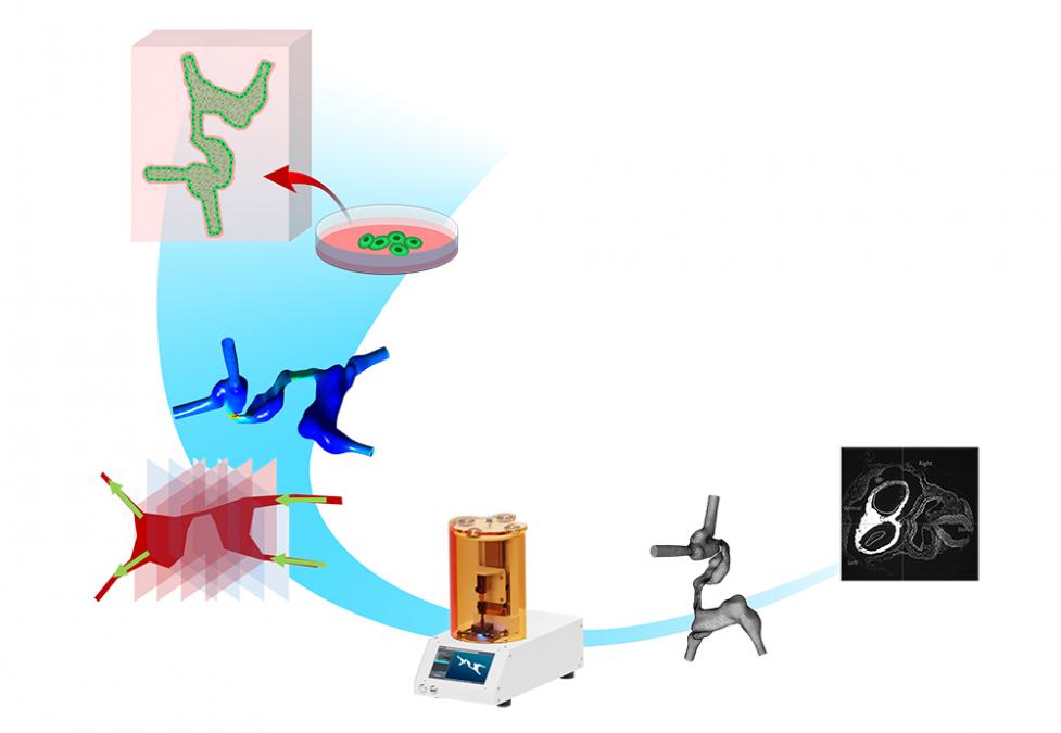
A diagram of the workflow in Vahid Serpooshan's National Science Foundation Early Career Development award study. The team starts with processing medical imaging data of patients to create 3D digital models of the developing human heart. These models are then bioprinted, seeded with cells, and analyzed using a variety of biomechanical and cellular assays to study the role of the tissue microenvironment in normal and abnormal processes of heart development. (Illustration Courtesy: Vahid Serpooshan)
“Up until this point, printing a synthetic, plastic model and perfusing it with different types of media has been done. But when it comes to hydrogels, and adding cells, and then having this flow going through — this is something that is a lot more complex, and no one has really tried this before,” said Serpooshan, assistant professor in the Wallace H. Coulter Department of Biomedical Engineering at Georgia Tech and Emory. “We bioprint with soft hydrogels that mimic heart tissue, and we add cells that look exactly like the real heart tissue cellular structure. Then we perfuse with the flow that mimics the developing heart flow.”
Nothing that exists now can replicate heart development in quite the same way, Serpooshan said. Animal models are imperfect substitutes for human heart development, and 2D models lack fidelity to the three-dimensional structures and flow at play.
“Having a bioprinted, engineered model that you can print hundreds of is one of our main advantages. You can order the machine to print hundreds of consistent models,” Serpooshan said. “This allows us to change parameters and study how cells behave without using any animals or even going to clinical trials.”
That means the models could be used to accurately test promising new drugs. They could also be used to help surgeons hone their techniques and develop new methods. Early versions of the models have been used by Serpooshan’s close collaborator Holly Bauser-Heaton in just that way. She’s a pediatric cardiologist at Emory.
In particular, Serpooshan and his team are focused on the velocity of blood flow through the developing heart in all three dimensions and the shear stresses that flow exerts on heart cells. Serpooshan said these are critical signals to cells that guide when they grow or move or change. When flow is altered, so, too, is the tissue development.
Serpooshan
“There is a theory called ‘no flow, no grow,’ that says that any disruption in the flow of the blood during the development of the heart could result in significant abnormal development,” he said. “That's where the significance of these measurements comes. Being able to visualize and quantify in 3D the flow parameters, including velocity and shear stress, helps us to study what cells are sensing in these environments and if we disrupt flow, for example, how that could change cell behavior.”
Of course, what Serpooshan is proposing is not easy. Heart tissue is complex, so creating this kind of model wasn’t even imaginable until 3D bioprinting came along, he said. The technique allows Serpooshan to deposit specific kinds of cells and biomaterials in specific areas of the tissue models to accurately reflect actual heart tissue composition.
They use three kinds of heart cells, created by reprogramming patients’ blood cells or skin cells into stem cells. This induced pluripotent stem cell technology allows Serpooshan to turn those stem cells into cardiac muscle cells and endothelial cells in the developing heart.
Building the models requires detailed imaging and processing to turn the scans into 3D structures before printing can begin. Serpooshan said other Coulter Department faculty, including Lakshmi “Prasad” Dasi, David Frakes, and Brooks Lindsey, as well as collaborators at Emory School of Medicine, including Bauser-Heaton and Timothy Slesnick, bring key expertise to the work.
Once his team has functioning models, they’ll be able to study two other key conditions along with blood flow that make up the microenvironment around cells as the heart forms and could affect their behavior: stiffness of the tissues and the concentrations of the different proteins in those tissues.
Data suggests all three contribute to abnormalities that lead to congenital heart defects. The question is, how significant is their role?
“Gaining knowledge about some very complex and vague processes that no one has been really able to study in such precision is going to be one of the main outcomes,” Serpooshan said.
Contact
Joshua Stewart
Communications Manager
Wallace H. Coulter Department of Biomedical Engineering
Latest BME News
Commercialization program in Coulter BME announces project teams who will receive support to get their research to market.

Courses in the Wallace H. Coulter Department of Biomedical Engineering are being reformatted to incorporate AI and machine learning so students are prepared for a data-driven biotech sector.

Influenced by her mother's journey in engineering, Sriya Surapaneni hopes to inspire other young women in the field.
Coulter BME Professor Earns Tenure, Eyes Future of Innovation in Health and Medicine
The grant will fund the development of cutting-edge technology that could detect colorectal cancer through a simple breath test
The surgical support device landed Coulter BME its 4th consecutive win for the College of Engineering competition.

New research from Georgia Tech helps doctors predict how therapies will interact with a child's immune system, potentially improving outcomes and reducing risks.