New technology can transform the way scientists view and interpret microscopic images
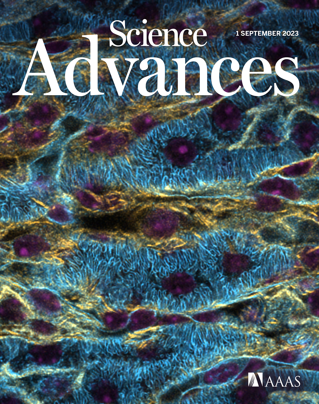
The cover of the September issue of Science Advances features a three-dimensional image of a kidney section acquired via laser scanning confocal microscopy. The three colors correspond to three fluorescent stains: DAPI (purple), phalloidin (blue), and wheat germ agglutinin (yellow). Fluorescence microscopy is an important tool in biological research, but the content and quality of acquired images limits observations of biological phenomena. New research from the lab of Shu Jia improves image quality and could transform the way scientists view and interpret microscopic images. Photo credit: © 2023 Shu Jia
A team of Georgia Tech and Emory researchers has introduced a revolutionary system to enhance the clarity and quality of fluorescence microscopy images. Their innovation, Multiscale Image Restoration through Optimally-Sparse Representation, or MIRO, could transform the way scientists view and interpret microscopic images.
“This has the potential to expand the horizons of biological knowledge,” said Shu Jia, professor in the Wallace H. Coulter Department of Biomedical Engineering at Georgia Tech and Emory, who led the research team. Their groundbreaking study is published this month as the cover story in the journal Science Advances.
Fluorescence microscopy stands as an unrivaled tool in biological research, illuminating the depths and functions of cellular structures. But Jia stressed the need for improved image clarity.
“The inherent limitations of current imaging detectors have posed significant challenges, with various noise sources clouding the resultant image,” he said.
Such noise impedes the precise observation of fluorescent signals, thereby restricting the full potential of fluorescence microscopy.
“The sensitivity discrepancies of current sensors coupled with pixel-to-pixel variations result in uncertain photon detection,” noted the study’s lead author, Biagio Mandracchia, a former postdoc in the Jia lab and currently an independent researcher at the University de Valladolid in Spain. “This has led to the production of images marred by noise, leaving researchers with suboptimal visual data for analysis."
A Clearer Future
The research team believes MIRO represents the future of image restoration.
“MIRO offers a comprehensive algorithm to conquer all noise sources pertinent to microscopy,” said Jia, whose collaborators include Shuichi Takayama, professor in the Coulter Department, as well as Shuyi Nie from Georgia Tech’s School of Biological Sciences, and Chunhui Xu, professor in the Department of Pediatrics at Emory’s School of Medicine.
“Unlike pre-existing methodologies that cater only to specific microscopy or camera types, MIRO boasts an adaptive, generalizable solution,” Jia added. “This robust approach encompasses a theoretical model that acknowledges all common noise types while extracting biological features from the surrounding noise.”
In practical terms, MIRO's capability was proven through its application across a diverse range of fluorescence microscopy techniques. The results displayed images of unparalleled clarity, enhancing both the observation and downstream analysis of specimens.
The research team also integrated MIRO with deep neural networks, a union of deterministic knowledge with machine intelligence that paves the way for rapid, high-quality image denoising.
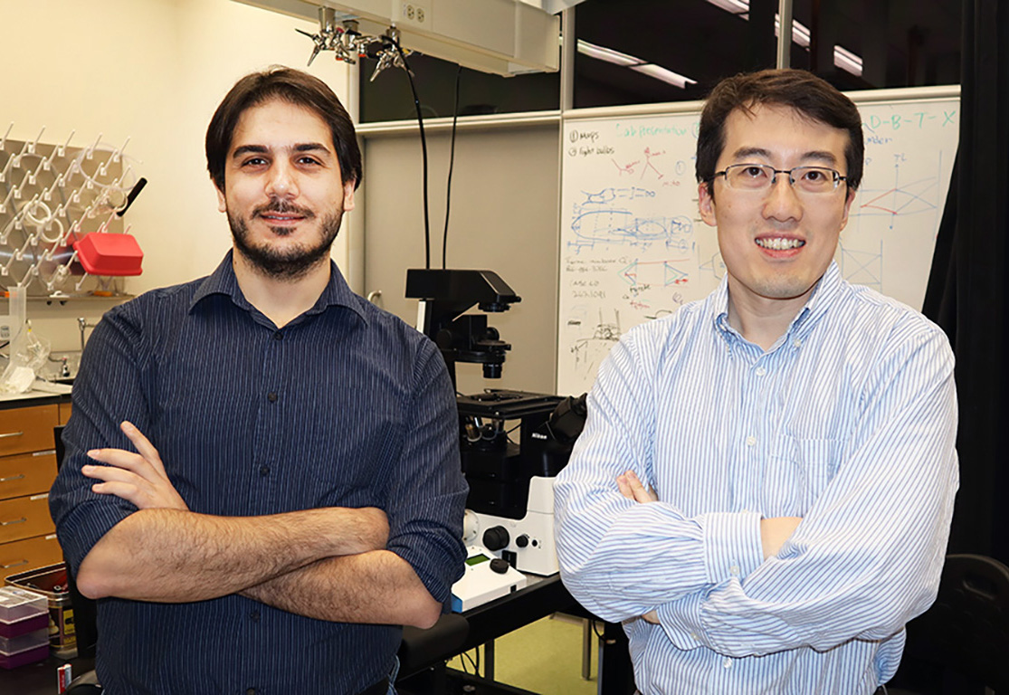
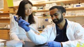
Researchers Biagio Mandracchia and Shu Jia led development of a new fluorescence microscopy system.
“This sets the stage for groundbreaking discoveries in imaging,” Jia said. “As scientists across the globe continue to push the boundaries in fluorescence microscopy, the introduction of MIRO ensures that the images captured will be clearer, more accurate, and immensely informative. This work not only enhances the quality of visual data but also magnifies the depth and breadth of understanding in biological research.”
The research was supported by the National Institutes of Health grants R35GM124846 and R01AA028527, the National Science Foundation grants BIO2145235 and EFMA1830941, and the Marvin H. and Nita S. Floyd Research Fund.
CITATION: Biagio Mandracchia, Wenhao Liu, Xuanwen Hua, Parvin Forghani, Soojung Lee, Jessica Hou, Shuyi Nie, Chunhui Xu, Shu Jia. “Optimal sparsity allows reliable system-aware restoration of fluorescence microscopy images.” Science Advances.
Latest BME News

Jo honored for his impact on science and mentorship
The department rises to the top in biomedical engineering programs for undergraduate education.

Commercialization program in Coulter BME announces project teams who will receive support to get their research to market.

Courses in the Wallace H. Coulter Department of Biomedical Engineering are being reformatted to incorporate AI and machine learning so students are prepared for a data-driven biotech sector.

Influenced by her mother's journey in engineering, Sriya Surapaneni hopes to inspire other young women in the field.
Coulter BME Professor Earns Tenure, Eyes Future of Innovation in Health and Medicine
The grant will fund the development of cutting-edge technology that could detect colorectal cancer through a simple breath test
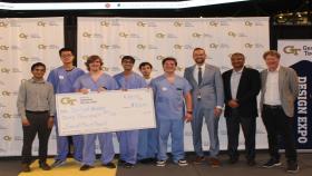
The surgical support device landed Coulter BME its 4th consecutive win for the College of Engineering competition.